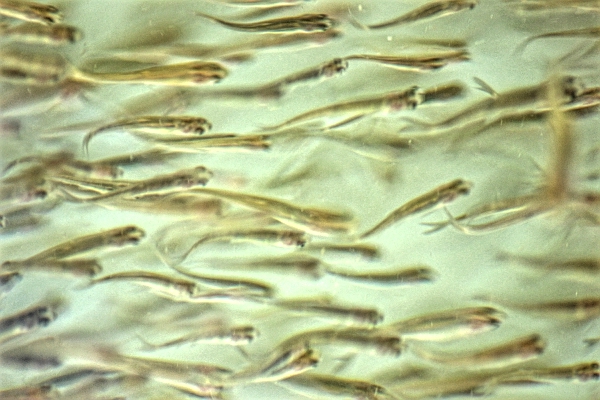
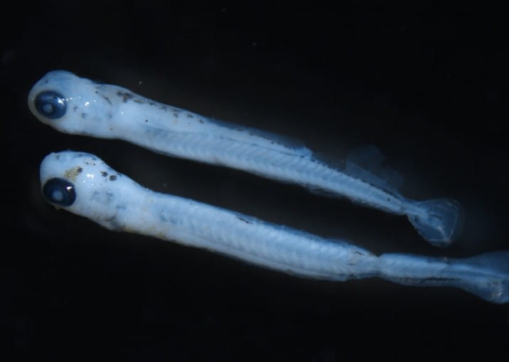
Two specimens of fathead minnow, each infected with a xenoma of
4.5 (523) · $ 19.00 · In stock

Download scientific diagram | —Two specimens of fathead minnow, each infected with a xenoma of the microsporidian Glugea pimephales protruding from the body near the anal vent (scale bar ¼ 6 mm). from publication: Occurrence of Glugea pimephales in Planktonic Larvae of Fathead Minnow in Algonquin Park, Ontario | The microsporidian Glugea pimephales was found parasitizing larval fathead minnow Pimephales promelas in Scott Lake, Algonquin Park, Ontario. These fish were estimated to be 2-3 weeks posthatch and, given the development time of the parasite, must have acquired infection soon | Glugea, Plankton and Larva | ResearchGate, the professional network for scientists.

Mean number of branchial xenomas observed in chinook and coho salmon
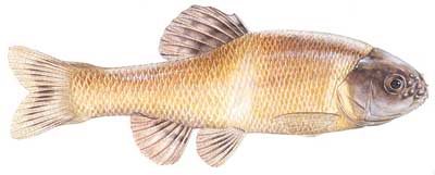
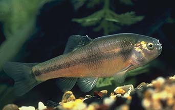
Fathead Minnow (Pimephales promelas)

Fathead minnow - Wikipedia
Fathead Minnow (Reptiles, Amphibians and Fish of the Kaibab National Forest) · iNaturalist

Fathead Minnow Mexican Fish.com

Efficacy of Intraperitoneally and Orally Administered ProVale, a Yeast β‐(1,3)/(1,6)‐D‐glucan Product, in Inhibiting Xenoma Formation by the Microsporidian Loma salmonae on Rainbow Trout Gills - Guselle - 2010 - North American Journal
Fathead Minnow Larval Fish Identification

Figure 1 from Migration, site selection, and development of Ornithodiplostomum sp. metacercariae (Digenea: Strigeoidea) in fathead minnows (Pimephales promelas).

5 Microsporidia

Aquaculture - Histological Change of Aquatic Animals by Parasitic Infection Watchariya Purivirojkul [1] Department of Zoology, Faculty of Science, Kasetsart University, Thailand 1. Introduction The aquatic parasite usually classified in six groups